EMG (Electromyography)
Electromyography (EMG) is a diagnostic test that measures the electrical activity of muscles and nerves. It is used to assess neuromuscular disorders, muscle weakness, and nerve dysfunction, such as in conditions like neuropathy, muscular dystrophy, and carpal tunnel syndrome. During the test, small electrodes or needle sensors are placed on or into the muscles to detect electrical signals during rest and movement. EMG helps doctors diagnose nerve or muscle damage, guiding treatment plans for various neurological and muscular conditions.
Electromyography (EMG): A Vital Diagnostic Tool for Neuromuscular Disorders
Electromyography (EMG) is a diagnostic test used to evaluate the electrical activity of muscles and the nerves that control them. It helps detect neuromuscular abnormalities by recording the electrical signals generated by muscle cells when they are activated. EMG is commonly used to diagnose conditions affecting the muscles and peripheral nerves, such as neuropathy, myopathy, muscular dystrophy, amyotrophic lateral sclerosis (ALS), and carpal tunnel syndrome. This test is essential for understanding the causes of muscle weakness, pain, numbness, and other symptoms related to nerve or muscle dysfunction.
How EMG Works
EMG measures the electrical activity in muscles at rest and during contraction. The procedure involves two main techniques:
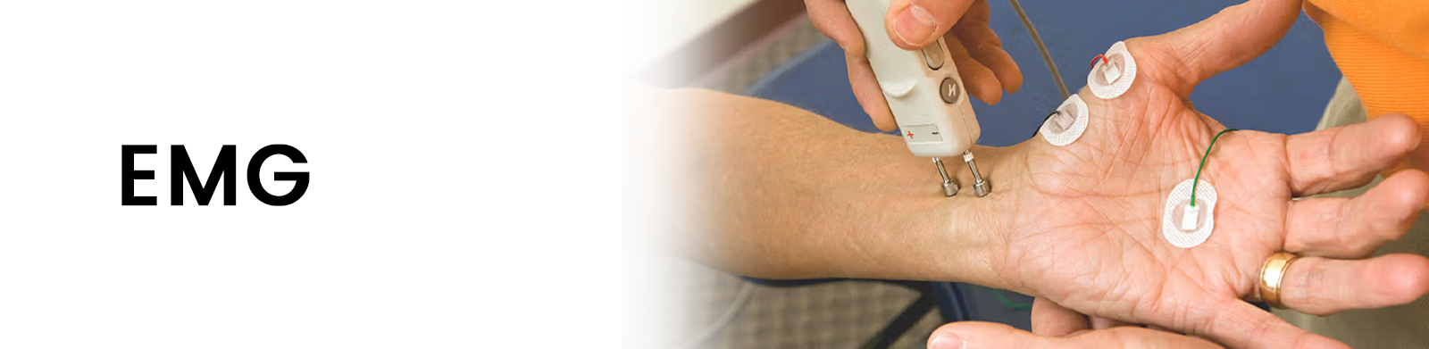
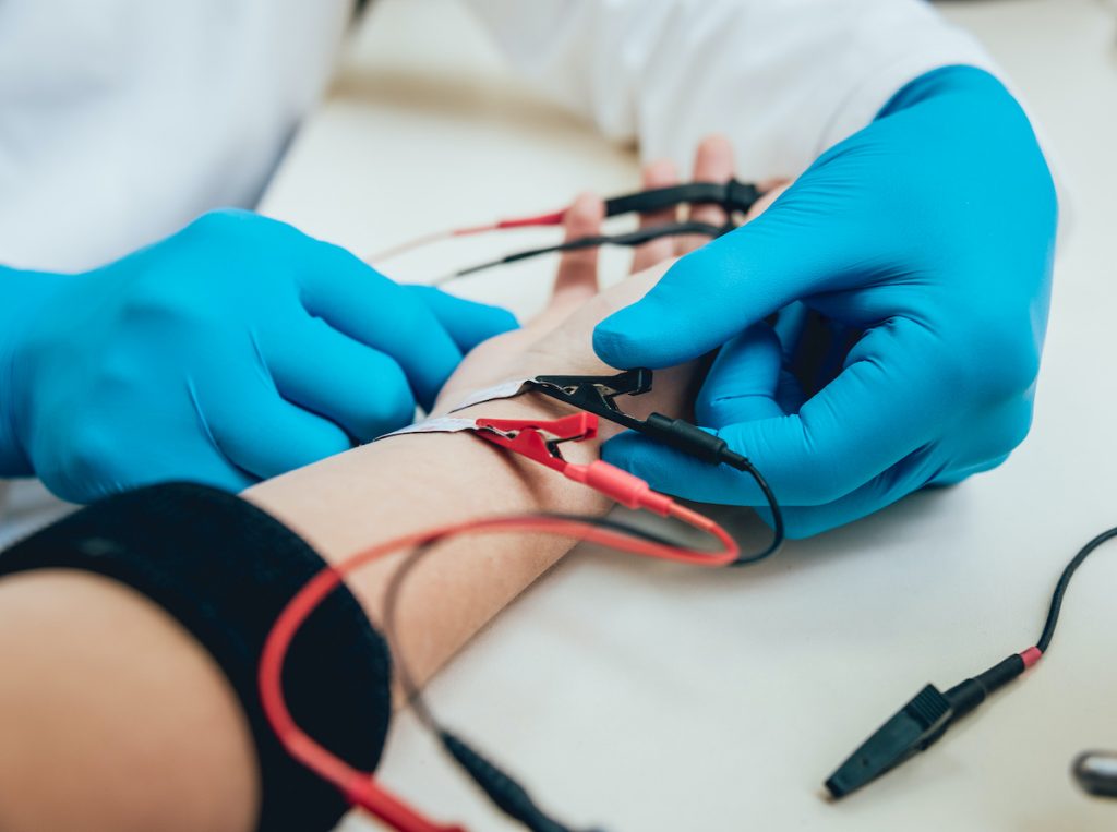
- Surface EMG (sEMG): Small adhesive electrodes are placed on the skin over the muscle being tested. This method is non-invasive and typically used for general muscle function assessments.
- Needle EMG: A thin, sterile needle electrode is inserted directly into the muscle to record electrical activity. This method provides more detailed and accurate information about muscle function and nerve signals.
The test records electrical impulses that appear as waveforms on a monitor. Normal muscle activity shows little to no electrical signal when at rest and a steady increase in activity during voluntary movement. In contrast, abnormal patterns—such as spontaneous muscle activity or irregular firing—can indicate nerve or muscle disorders.
Applications of EMG
EMG is widely used in neurology, orthopedics, and rehabilitation medicine to diagnose and monitor a range of conditions, including:
1. Nerve Disorders (Neuropathies)
EMG helps detect peripheral neuropathies, such as diabetic neuropathy and carpal tunnel syndrome, which result from nerve compression or damage. Patients experiencing numbness, tingling, or weakness in the hands and feet often undergo EMG to determine the severity and location of nerve dysfunction.
2. Muscle Disorders (Myopathies)
Muscle diseases like muscular dystrophy, polymyositis, and myasthenia gravis affect muscle strength and function. EMG identifies abnormal muscle activity, helping differentiate between muscle and nerve-related conditions.
3. Motor Neuron Diseases
EMG is crucial in diagnosing amyotrophic lateral sclerosis (ALS) and other motor neuron diseases. These conditions cause progressive muscle weakness and atrophy due to nerve degeneration. EMG findings, such as fasciculations (muscle twitches) and spontaneous discharges, can indicate nerve cell damage.
4. Spinal and Radiculopathy Conditions
Patients with herniated discs, sciatica, or spinal nerve compression may experience pain, weakness, or numbness in the limbs. EMG, combined with nerve conduction studies (NCS), helps determine if symptoms are due to nerve root irritation or muscle dysfunction.
Advantages and Limitations of EMG
Advantages:
- Accurately identifies nerve and muscle disorders
- Helps differentiate between neurological and muscular conditions
- Provides real-time data for clinical diagnosis and treatment planning
- Useful in monitoring disease progression and recovery
Limitations:
- Needle EMG can be mildly uncomfortable or painful
- Results may vary based on examiner expertise
- Cannot detect central nervous system disorders like stroke or multiple sclerosis
Conclusion
Electromyography (EMG) is a vital diagnostic tool in neurology and rehabilitation, helping to detect and assess nerve and muscle disorders. By analyzing electrical activity in muscles, EMG provides critical insights into the function of the neuromuscular system. Though it has some limitations, its accuracy and clinical significance make it an indispensable test for diagnosing and managing a wide range of neuromuscular conditions.
FAQs
1. What is an EMG?
EMG (Electromyography) is a diagnostic test that measures muscle activity and nerve function to detect neuromuscular disorders.
2. How does an EMG work?
A small needle electrode is inserted into a muscle to record electrical signals, helping doctors evaluate muscle and nerve function.
3. Why is an EMG performed?
An EMG helps diagnose nerve and muscle disorders, including neuropathy, myopathy, ALS, carpal tunnel syndrome, and radiculopathy.
4. Is an EMG painful?
The test may cause mild discomfort as small needles are inserted into muscles, but it is generally tolerable.
5. How long does an EMG take?
The test usually lasts 30 to 60 minutes, depending on the number of muscles and nerves being tested.